

Extreme cold challenges the human body’s ability to maintain a stable internal environment. The core temperature is normally held near 37 degrees Celsius through a combination of behavioral choices and automatic physiological processes. When heat loss exceeds heat production and conservation for an extended period, core temperature falls and a series of predictable yet increasingly dangerous changes occur. These changes affect every major organ system, damage tissues directly, and can culminate in death or permanent disability. Understanding the sequence of events helps explain why rapid intervention and proper preparation matter so much in cold environments, whether on land or in water.
The body detects cooling through thermoreceptors in the skin and deeper tissues. Signals reach the hypothalamus, which coordinates responses aimed at preserving heat for the brain, heart, and other vital organs. The first and often most effective actions are behavioral: seeking shelter, adding clothing layers, or increasing physical activity. When these are insufficient or impossible, automatic mechanisms take over.
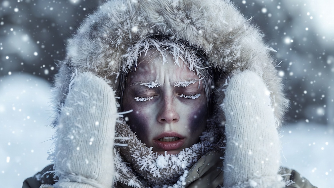
Peripheral vasoconstriction is the primary physiological defense. Sympathetic nerves cause blood vessels in the skin and extremities to narrow sharply. Normal skin blood flow of roughly 250 milliliters per minute can fall below 50 milliliters per minute and, in extreme cases, approach zero. This reduces convective and radiative heat loss from the body surface while increasing overall insulation. Skin temperature drops quickly, sometimes approaching the temperature of the surrounding air or water, while core temperature is defended. The trade-off is clear: extremities become cold, stiff, and numb, raising the risk of localized injury. Insulation improves markedly once skin temperature falls below about 35 degrees Celsius and reaches maximum when skin temperature drops to 31 degrees Celsius or lower.
Piloerection, or goosebumps, occurs when tiny muscles at hair follicles contract. This traps a thin layer of still air next to the skin, but the effect is modest in humans because of limited body hair. More important is the metabolic response. When core or skin temperature continues to decline, shivering begins. This involuntary, rhythmic muscle activity can increase heat production two to five times above resting levels. Shivering usually starts in the torso and spreads outward. It relies on carbohydrate oxidation initially and draws heavily on muscle glycogen stores. Cardiac output rises mainly through increased stroke volume rather than heart rate. In young adults, oxygen consumption during strong shivering can reach 1 to 2 liters per minute depending on conditions. Non-shivering thermogenesis through brown adipose tissue plays only a minor role in adults, unlike in infants.
If heat loss continues to outpace these defenses, core temperature falls below 35 degrees Celsius. This defines hypothermia, a medical emergency that progresses through recognizable stages. In mild hypothermia (roughly 32 to 35 degrees Celsius), intense shivering persists. The person feels cold, fatigued, and uncoordinated. Confusion appears early because the brain is sensitive to even small temperature drops. Speech may slur, judgment falters, and some individuals experience a paradoxical sensation of warmth that can lead them to remove clothing. Skin looks pale as blood is shunted inward. Breathing becomes rapid and shallow. At this stage the body is still fighting, but impaired decision-making increases danger.
Moderate hypothermia (approximately 28 to 32 degrees Celsius) brings more ominous changes. Shivering weakens and eventually stops as muscle energy stores deplete and neural drive declines. The person grows drowsy, apathetic, or markedly confused. Movements become slow and clumsy. Pulse slows (bradycardia), blood pressure falls, and breathing rate decreases. Skin may turn blue or appear puffy in exposed areas. Amnesia or irrational behavior is common. Cardiac irritability rises, setting the stage for dangerous arrhythmias. Blood viscosity increases because of fluid shifts and cold-induced changes in blood components, raising the risk of clotting abnormalities.
Severe hypothermia occurs below 28 degrees Celsius. The victim loses consciousness. Shivering is absent. Muscles become rigid. Pupils dilate and become unresponsive. Breathing slows dramatically or ceases. Heart rhythm destabilizes, often progressing to ventricular fibrillation or asystole. Cardiac output and blood pressure drop to levels that cannot sustain life. Metabolic rate falls overall, which paradoxically reduces oxygen demand and can allow brief survival at very low temperatures, but without external rewarming and advanced life support the outcome is almost always fatal. Multiple systems fail together: the brain’s electrical activity slows, kidney function shifts toward cold diuresis (increased urine output from reduced antidiuretic hormone and central blood volume changes), and immune responses weaken.
Even when core temperature remains above the hypothermic range, localized tissues can freeze. Frostbite develops once skin and underlying tissue temperatures drop below 0 degrees Celsius. Intense vasoconstriction first reduces blood flow dramatically. Below freezing, ice crystals form initially in the extracellular space, drawing water out of cells and causing dehydration and electrolyte imbalance. Continued exposure allows intracellular ice formation that ruptures cell membranes and organelles. Vascular damage leads to stasis, microthrombus formation, and ischemia. On rewarming, reperfusion injury adds inflammation, cytokine release, and further tissue destruction.
Clinically, frostbite is often staged similarly to burns. First-degree injury shows numbness, pallor, and surrounding redness with later peeling. Second-degree produces clear blisters after rewarming. Third-degree involves hemorrhagic blisters and full-thickness skin loss. Fourth-degree extends into muscle, tendon, or bone, frequently requiring amputation. The most vulnerable sites are fingers, toes, nose, ears, and cheeks because of high surface-area-to-volume ratios and dependence on peripheral blood flow. Wind dramatically accelerates heat loss through convection, while moisture increases conductive loss. Risk factors include tight or wet clothing, impaired circulation from smoking or vascular disease, alcohol use (which causes vasodilation), malnutrition, and extremes of age.
Not all cold injuries involve actual freezing. Non-freezing cold injuries occur after prolonged exposure to wet conditions above freezing, typically between 0 and 10 degrees Celsius. Trench foot, also called immersion foot, results from sustained vasoconstriction combined with moisture that macerates skin and damages nerves and small vessels. The foot first becomes pale, numb, cold, and clammy. On rewarming, intense pain, redness, swelling, and sometimes blistering appear. Long-term consequences often include chronic neuropathic pain, cold hypersensitivity, and increased susceptibility to future injury. Chilblains (pernio) arise from repeated damp, non-freezing cold exposure. They present as itchy, red or purple swollen patches caused by an inflammatory response to vascular leakage and endothelial damage. Both conditions highlight that cold injury does not require ice crystal formation; ischemia and inflammation alone can produce lasting harm.
Cold water creates a far more hostile environment than air at the same temperature. Water conducts heat away roughly 25 times faster than air, and convective losses can be even greater. The sequence of events in cold-water immersion follows a predictable pattern. In the first one to three minutes, cold shock triggers an uncontrollable gasp reflex, hyperventilation, and panic. This phase alone can cause drowning before hypothermia develops. Over the next 3 to 30 minutes, muscle and nerve cooling produce swim failure: dexterity is lost, grip strength collapses, and coordinated swimming becomes impossible. Only after this incapacitation phase does core temperature fall significantly. Survival time estimates vary widely with water temperature, body size, clothing, and flotation, but in near-freezing water without protection, useful consciousness is often measured in minutes rather than hours. Many published charts focus on hypothermia timelines while understating the early risk of drowning from cold shock or swim failure.
Individual factors strongly influence outcomes. Subcutaneous fat provides insulation and reduces the shivering required to maintain temperature. Greater muscle mass supports higher heat production. Older adults often show slower vasoconstriction and reduced shivering capacity because of lower muscle mass and blunted sympathetic responses. Women, on average, have a higher surface-area-to-mass ratio and less lean mass for heat generation, leading to faster core cooling in water even when fat thickness is similar. Acclimatization through repeated cold exposure can enhance metabolic responses or improve vascular control in some individuals, although results vary. Proper layered clothing that traps air, blocks wind, and stays dry remains the single most effective protection. Wet clothing or immersion eliminates trapped air and multiplies heat loss. Alcohol and certain drugs impair judgment and blunt protective vasoconstriction. Pre-existing conditions such as diabetes, heart disease, or peripheral vascular disease accelerate injury.
Survival at extremely low core temperatures has been documented in rare cases, illustrating both the body’s resilience and the importance of rapid, careful rewarming. One of the lowest recorded core temperatures in an adult survivor of accidental hypothermia reached 13.7 degrees Celsius after prolonged immersion in icy water followed by prompt resuscitation and extracorporeal rewarming. Children have survived even lower temperatures in some documented instances. These cases succeed partly because profound hypothermia lowers metabolic demand and protects the brain from ischemic damage during the period of low or absent circulation. However, after rescue an “afterdrop” phenomenon can occur: as peripheral vessels dilate during rewarming, cold blood returning from the limbs can further lower core temperature and precipitate collapse or arrhythmias.
Survivors of significant cold exposure may face lasting consequences. Deep frostbite often results in tissue loss requiring amputation and chronic pain or sensitivity in remaining areas. Non-freezing injuries frequently produce long-term neuropathic pain and cold intolerance that can persist for years. Severe hypothermia can leave residual cognitive deficits, cardiac conduction abnormalities, or kidney impairment, although many patients recover fully with modern care. Repeated or prolonged cold stress may also increase susceptibility to future injury through vascular or nerve damage.
The human body possesses sophisticated mechanisms to defend core temperature, yet these defenses have clear limits. Vasoconstriction and shivering buy time, but without external help or escape from the cold environment they eventually fail. Localized freezing or prolonged wet cold destroys tissue even when the core remains protected. In water the timeline compresses dramatically because of rapid heat loss and early incapacitation. Knowledge of these processes reveals why prevention through appropriate clothing, awareness of conditions, and avoidance of alcohol or fatigue in cold settings is far more effective than any treatment after injury has occurred. Extreme cold does not negotiate; it simply removes heat until the body can no longer compensate. Respect for that reality remains the most reliable safeguard.