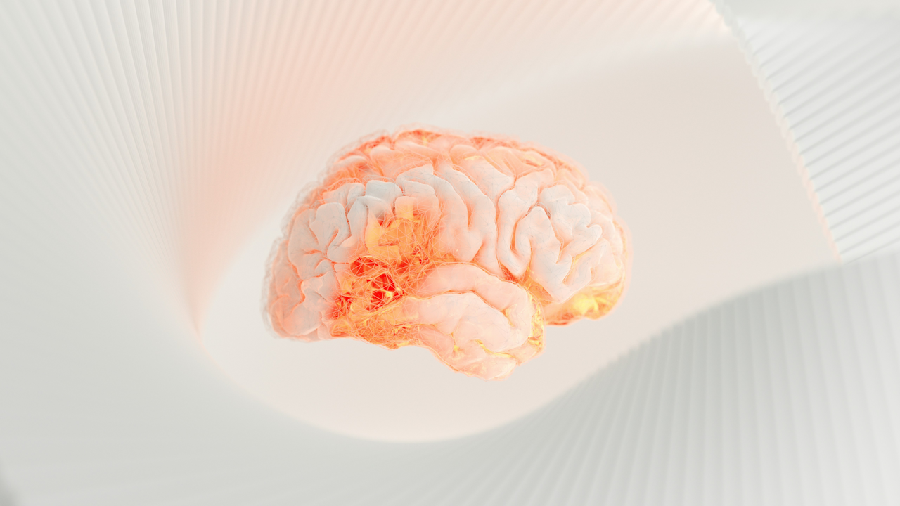
Memory is one of the most fascinating aspects of human cognition. It allows us to learn from experiences, build relationships, and navigate the world. At its core, memory involves the brain’s ability to encode, store, and retrieve information. However, memories are not permanent records etched into our minds like data on a hard drive. They are dynamic, changeable, and sometimes even erasable. This article explores the intricate processes behind how the brain forms memories and how it erases or forgets them, drawing on recent neuroscience research. We will delve into the brain regions involved, the cellular mechanisms at play, and the implications for mental health and therapy.
The Basics of Memory Formation
Memory formation begins with encoding, the process of transforming sensory experiences into a form that the brain can store. When we encounter something new, like a conversation or a visual scene, our senses send signals to the brain. These signals trigger a cascade of neural activity. The hippocampus, a seahorse-shaped structure in the medial temporal lobe, plays a central role in this process. It acts as a hub for converting short-term memories into long-term ones, essentially binding different elements of an experience together.
Encoding involves several stages. First, sensory information is held in short-term or working memory, which has a limited capacity and lasts only seconds to minutes. For instance, remembering a phone number long enough to dial it relies on this system. If the information is deemed important, it moves to consolidation, where it is stabilized and integrated into long-term storage. Consolidation can occur over hours, days, or even longer, often strengthened during sleep. During this phase, the brain replays neural patterns from the day, reinforcing connections.
Long-term memories are categorized into types. Declarative memories include facts (semantic) and personal events (episodic), while procedural memories involve skills like riding a bike. The neocortex, the outer layer of the brain, stores these long-term memories, with different regions handling specific types. For example, the amygdala, an almond-shaped structure, adds emotional weight to memories, making fearful or joyful experiences more vivid.
At the cellular level, memory formation relies on synaptic plasticity, the ability of connections between neurons (synapses) to strengthen or weaken. When we learn, synapses create new circuits among the brain’s 100 billion neurons, each capable of forming up to 10,000 connections. Repeated exposure to an event strengthens these synapses through a process involving neurotransmitters like glutamate. Glutamate binds to receptors on neurons, enhancing signal transmission and making the memory pathway more efficient. For traumatic events, this can lead to an increase in glutamate receptors in the amygdala, encoding strong fear responses.
Research shows that the more we practice or revisit an experience, the stronger the synaptic connections become. A golfer perfecting a swing through repetition is essentially rewiring their brain’s motor circuits. Conversely, infrequent exposure weakens these links, leading to forgetting. This plasticity is key to learning but also explains why memories can fade over time.
The Physical Basis of Memories: Engrams
The concept of an engram provides a deeper understanding of memory’s physical foundation. An engram is a specific group of neurons that fire together to encode and recall a memory. This idea, first proposed over a century ago, was experimentally confirmed in animal models. For example, activating an engram in rats can trigger the recall of a fear-inducing event, proving that these neuronal clusters store memory traces.
Engrams form through unique firing patterns during an experience. When we encounter a familiar place, “place cells” in the hippocampus activate, contributing to the engram. Advanced imaging techniques now allow scientists to observe these cells in real time, revealing how memories are etched into neural networks. Engrams are not static; they can be modified or linked to new information, highlighting memory’s malleability.
Linking Events Over Time to Form Coherent Memories
Real-life memories often involve sequences of events separated by time. How does the brain connect a neutral sound to a later unpleasant sensation? Studies in mice reveal that the hippocampus bridges these temporal gaps through unexpected mechanisms. Rather than maintaining constant neural activity during delays, the brain uses sparse, burst-like patterns. These bursts efficiently encode associations, possibly by altering synaptic strengths without draining energy.
In experiments, mice learned to associate a tone with an air puff after a 15-second interval. Neural activity during the wait appeared random but formed complex patterns that linked the events. This “time-bridging” process is crucial for forming episodic memories and may underlie disorders like PTSD, where neutral cues trigger fear.
Specialized Cells in Memory Organization
Recent research has identified specific cell types that organize memories by timing. In the human brain, “boundary cells” and “event cells” help separate experiences into discrete units. Boundary cells activate during both subtle (soft) and major (hard) shifts in context, like a scene change in a video. Event cells respond only to hard boundaries, such as switching from a game to a commercial.
New memories form when both cell types peak, particularly after hard boundaries. This organization is like grouping photos into albums on a device: boundaries create new albums, while soft changes add to existing ones. For retrieval, the brain uses these markers to navigate memories efficiently. Participants in studies remembered items better after boundaries but struggled with order across hard ones, confirming separate storage.
These findings, from epilepsy patients with implanted electrodes, suggest potential therapies targeting dopamine neurons or brain rhythms like theta oscillations, which aid memory formation.
Memories Beyond the Brain: A New Frontier
Traditionally, memory is seen as a brain-exclusive function. However, emerging research shows that non-brain cells can also “remember” patterns. In experiments, nerve and kidney cells exposed to spaced chemical signals activated a “memory gene” more robustly than with continuous signals. This mirrors the “spaced learning” effect in brains, where breaks improve retention.
The same gene turns on in brain cells during pattern detection. This discovery implies that cells throughout the body, like those in the pancreas or even cancer cells, might retain information about past events, such as meal times or treatments. It opens new ways to study memory and could influence therapies for learning disorders.
The Process of Erasing or Forgetting Memories
Forgetting is not just a failure of memory; it is an active process essential for mental health. Without it, our minds would be overwhelmed by irrelevant details. The brain erases memories through several mechanisms, including intrinsic forgetting, where chronic signals degrade memory traces.
One key pathway involves “forgetting cells,” such as dopamine neurons that release signals onto engram cells. In fruit flies, this activates a cascade involving proteins like Rac1, PAK3, and cofilin, altering the actin cytoskeleton and dismantling synaptic structures. Rac1-dependent forgetting affects various memory types, while Cdc42 targets others. Neurogenesis in the hippocampus adds new neurons, remodeling circuits and clearing old traces.
Humans can consciously remove unwanted memories by dampening the circuits that store them. In studies, participants forgot specific items by reducing neural excitability in perceptual areas, making sensory channels less sensitive. This “hijacked adaptation” model uses top-down control to lower circuit gain, evidenced by EEG patterns like traveling waves. Such active removal helps manage intrusive thoughts and clears working memory space.
Hiding and Erasing Traumatic Memories
Traumatic memories are stored differently, often hidden from conscious access. During stress, extra-synaptic GABA receptors shift encoding, using pathways independent of glutamate. These memories reside in subcortical regions and require the same brain state for recall, as shown in mice given drugs mimicking stress. This “state-dependent” storage protects against overload but can contribute to PTSD or anxiety.
Scientists can now alter or erase memories by exploiting reconsolidation. When a memory is recalled, it becomes vulnerable for about 10 minutes. Showing cues without the original stimulus can update it, erasing fear responses. Optogenetics in animals allows precise manipulation of engram cells with light, even implanting false memories. These techniques hold promise for treating addiction, depression, and trauma.
Implications for Health and Future Therapies
Understanding memory formation and erasure has profound implications. For disorders like Alzheimer’s, enhancing synaptic plasticity could preserve memories. Drugs targeting glutamate receptors are under study to boost learning in cognitive impairments.
In mental health, erasing harmful memories could alleviate PTSD. Modulating forgetting cells or using behavioral interventions during reconsolidation windows might reduce rumination or hallucinations. However, ethical concerns arise: altering memories risks changing personal identity.
Future research will explore non-brain memory for holistic treatments, perhaps influencing how cancer cells “remember” therapies. As our knowledge grows, so does the potential to harness memory’s power for better lives.
In summary, the brain forms memories through intricate neural networks, engrams, and synaptic changes, primarily in regions like the hippocampus. It erases them actively via forgetting cells, dampening circuits, and reconsolidation. This duality ensures adaptability, but imbalances can lead to disorders. Ongoing discoveries continue to unravel these mysteries, promising innovative interventions.

